Our Technology
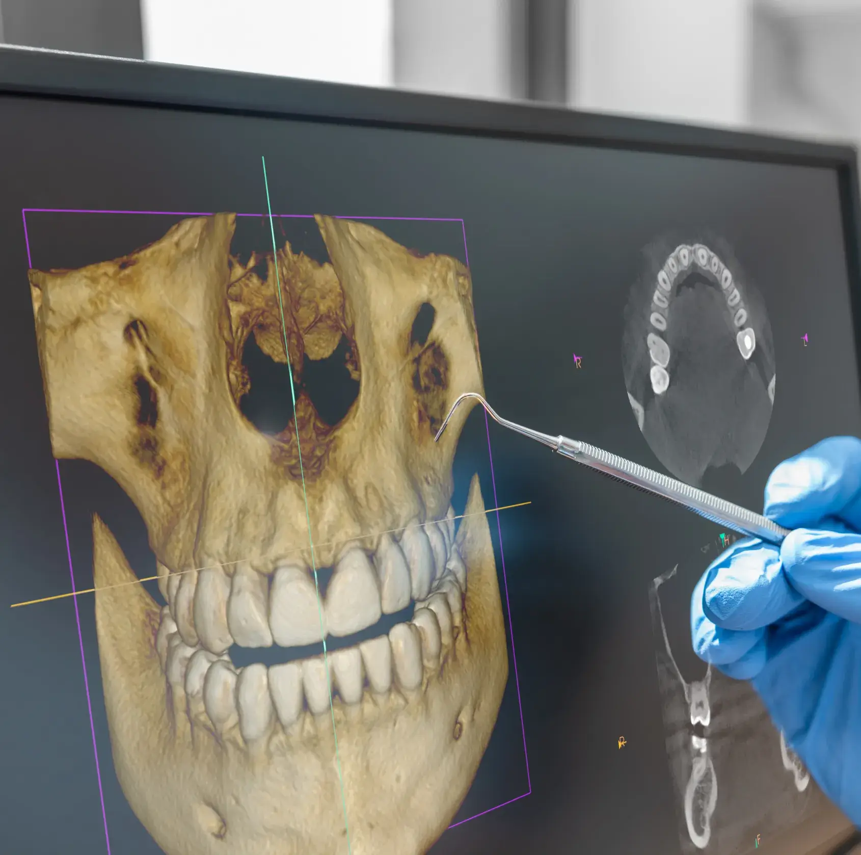
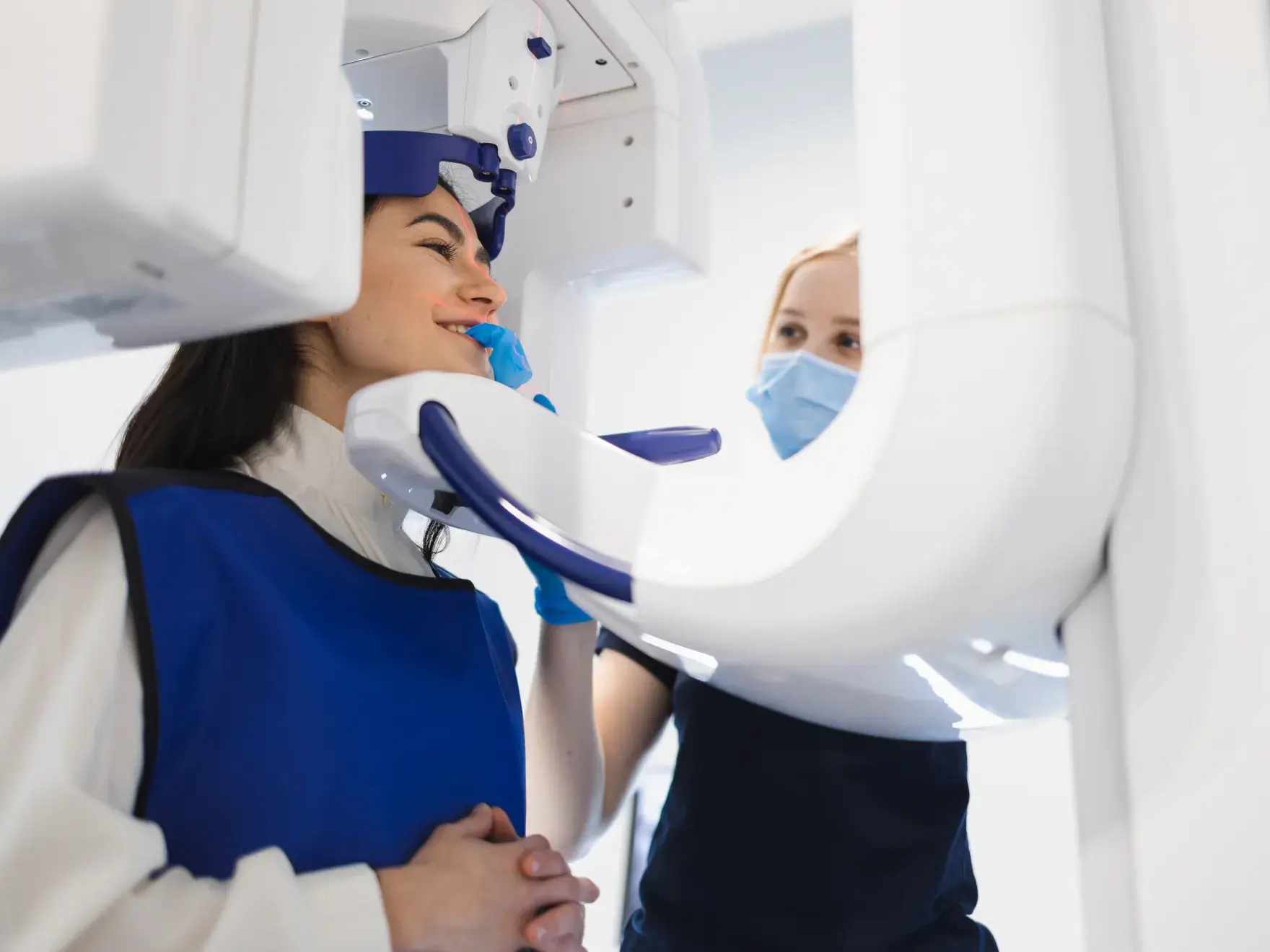
Cone Beam CT Imaging
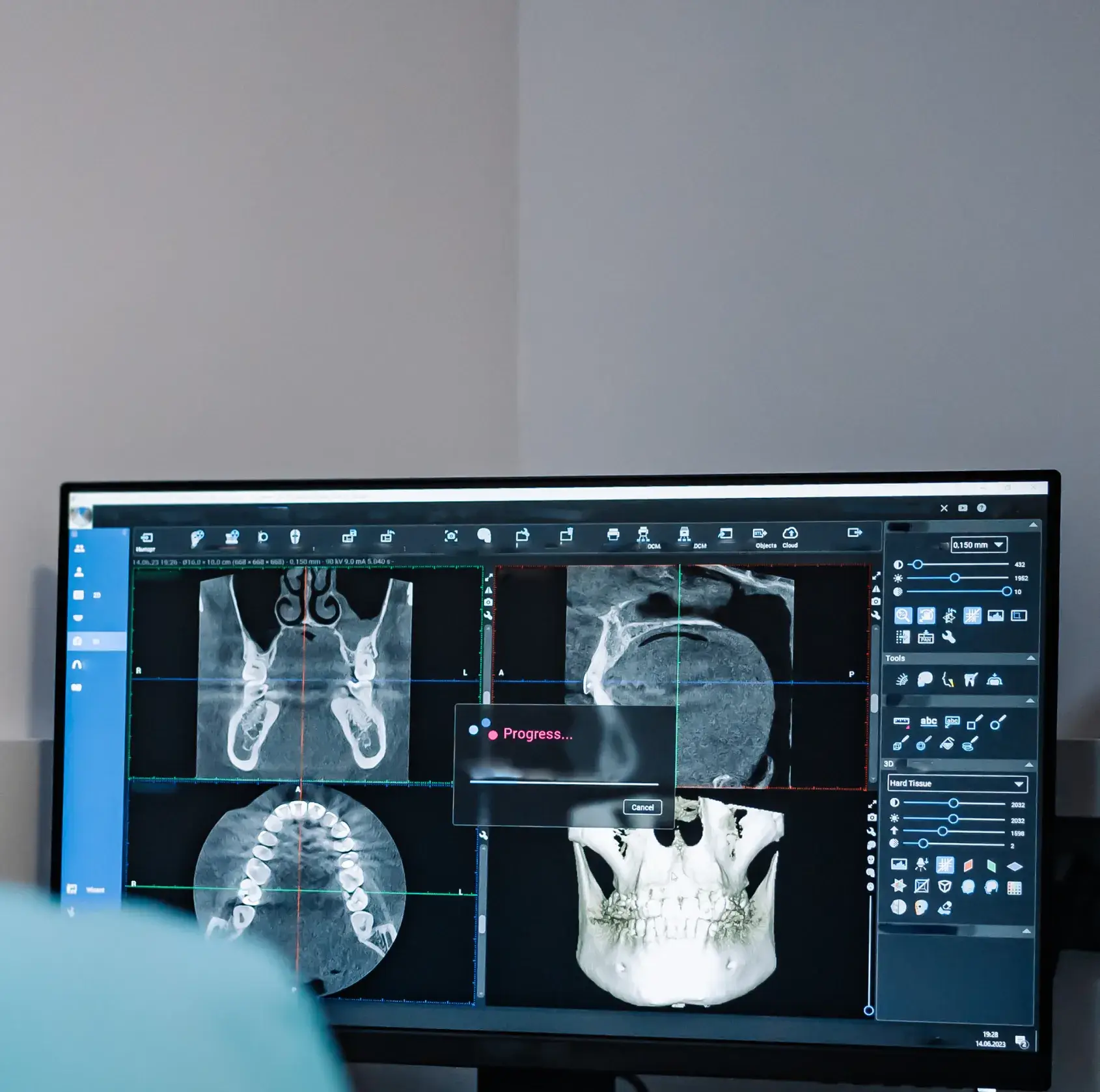
CBCT imaging produces a complete three-dimensional view of your teeth, jaw, bone structures, nerve pathways, and sinus anatomy in a single scan. The diagnostic detail it provides is not achievable with conventional two-dimensional X-rays.
Dr. Olivero uses CBCT imaging to:
- Evaluate bone density and volume at implant sites
- Identify the precise location of nerve pathways before surgical procedures
- Assess sinus anatomy in cases involving upper arch treatment
- Plan complex extractions and bone grafting with spatial accuracy
- Detect structural concerns that do not appear on standard X-rays
- Evaluate joint anatomy to help identify and diagnose TMD
- Analyze airway volume and cross-sectional area to help diagnose or screen for obstructive sleep apnea (OSA) and other respiratory restrictions.
Every finding is reviewed with you directly before any decisions are made.
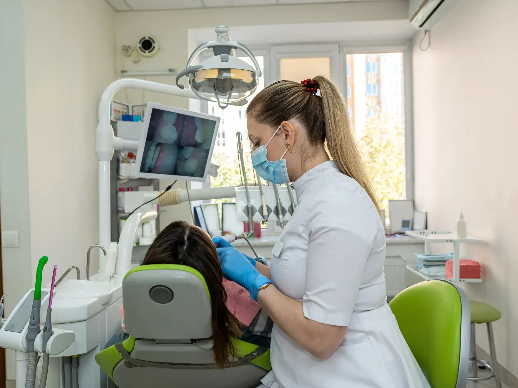
Intraoral Cameras
Intraoral cameras capture high-definition images of your teeth and gum tissue in real time. During your exam, Dr. Olivero uses these images to show you exactly what she sees, providing the visual clarity that transforms a clinical recommendation into a shared understanding.
Intraoral cameras allow Dr. Olivero to:
- Document the current condition of every tooth and surrounding tissue
- Show you areas of concern before treatment is discussed
- Track changes in your oral health across appointments over time
- Support fully informed decision-making at every visit
Digital X-Rays
Digital X-rays produce precise, high-resolution images immediately and with significantly less radiation than conventional film. Results are available within seconds, which means your diagnosis and treatment discussion happen within the same appointment.
Dr. Olivero uses digital X-rays to:
- Detect decay between teeth not visible during a clinical exam
- Monitor bone levels and identify early signs of bone loss
- Assess root health and the integrity of existing restorations
- Track changes in bone and tissue across recall visits over time
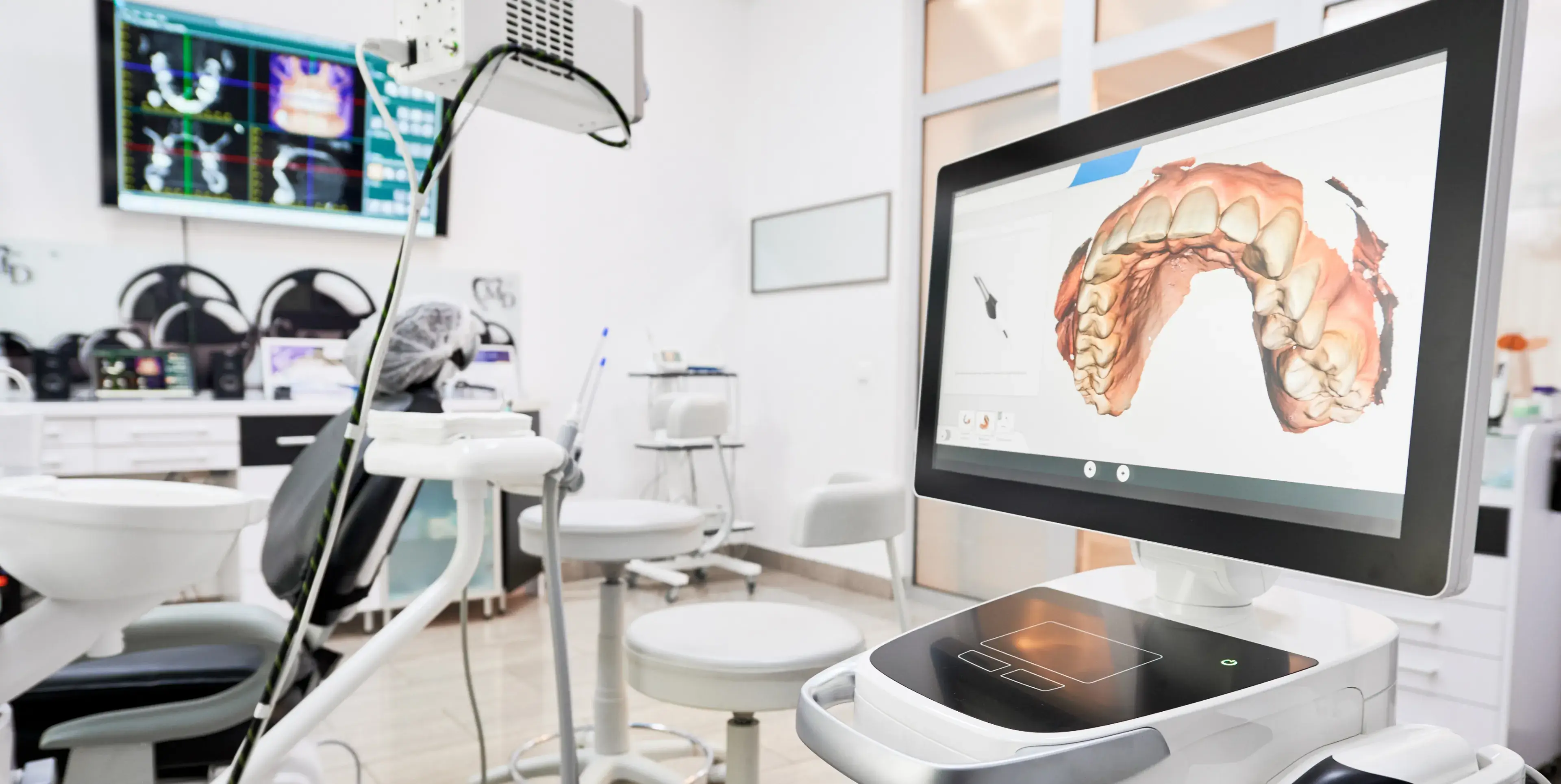
Digital Impressions
O Dental uses an intraoral scanner to capture precise digital models of your teeth, replacing traditional putty trays entirely. The scanner produces a highly accurate three-dimensional model of your dentition in minutes.
Digital impressions benefit patients by:
- Eliminating the discomfort of traditional impression trays and material
- Producing a more dimensionally accurate model than putty impressions
- Reducing the likelihood of remakes or ill-fitting restorations
- Shortening overall treatment time by streamlining the fabrication process
Crowns, bridges, dentures, and other restorations all benefit from the precision a digital impression provides.
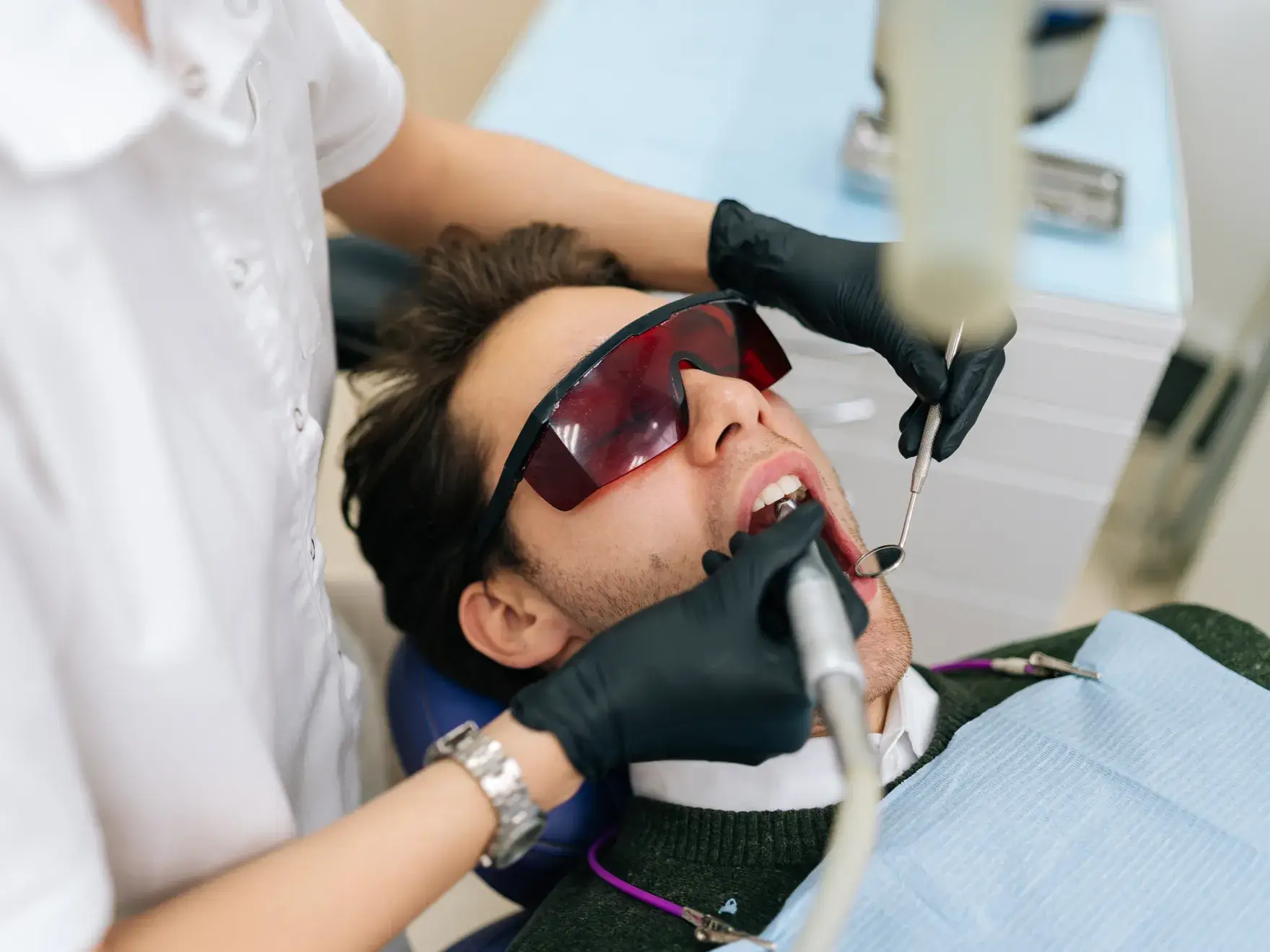
Laser Periodontal Therapy
For patients with periodontal disease, laser therapy provides a clinically effective alternative to traditional surgical intervention. The laser selectively removes diseased tissue while preserving the healthy gum tissue surrounding it.
Compared to conventional periodontal surgery, laser therapy offers:
- Greater precision in targeting diseased tissue
- Reduced procedural discomfort during treatment
- Less post-operative sensitivity and swelling
- Faster healing and recovery
- Minimal impact on healthy surrounding tissue